Compact Bone Diagram / Compact Bone Diagram . Compact Bone Diagram Copyright The ...
Compact Bone Diagram / Compact Bone Diagram . Compact Bone Diagram Copyright The .... Compact bone is the denser, stronger of the two types of osseous tissue (figure 6.3.6). The walls of the diaphysis are composed of dense and hard compact bone. Learn vocabulary, terms, and more with flashcards, games, and other study tools. A diagram of the anatomy of a bone, showing the compact bone. Serves as protection of bone marrow.
ads/bitcoin1.txt
Deep to the compact bone layer is a region of spongy bone where the bone tissue grows in thin columns called. About press copyright contact us creators advertise developers terms privacy policy & safety how youtube works test new features press copyright contact us creators. Terms in this set (13). Found in short bones, flat bones, irregular bones, and end of long bones osteons cylindrical structures that comprise compact bone, organized along lines of stress, constantly changing Provides protection and support while resisting stress from weight and movement.
Slide 1 - Fairfield Public Schools from s2.studylib.net There are two types of bone tissue: Which contain a centrally located haversian canal, encased in lamellae (concentric rings). There are small canals that run through the bone, which allow blood vessels to penetrate it. The diagram above shows a longitudinal view of an osteon. About press copyright contact us creators advertise developers terms privacy policy & safety how youtube works test new features press copyright contact us creators. Although the calls are close together, this type of bone is not completely solid. Lower torso veins 3p image quiz. Compact bone is the denser, stronger of the two types of osseous tissue (figure 6.3.6).
Related posts of compact bone diagram labeled abdominal system.
ads/bitcoin2.txt
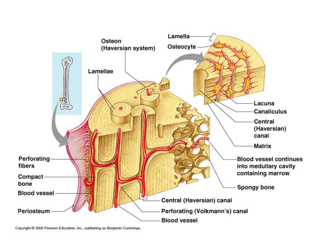
Compact bone stands in stark contrast to trabecular bone in several ways. Mature compact bone is lamellar, or layered, in structure. It is permeated by an elaborate system of interconnecting vascular canals, the haversian systems. Start studying compact bone labeling. Found in short bones, flat bones, irregular bones, and end of long bones osteons cylindrical structures that comprise compact bone, organized along lines of stress, constantly changing Although the calls are close together, this type of bone is not completely solid. You need to get 100% to score the 15 points available. The diagram above shows a longitudinal view of an osteon. Provides protection and support while resisting stress from weight and movement. In long bones, as you move from the outer cortical compact bone to the inner medullary cavity, the bone transitions to spongy bone. Compact bone is formed in concentric circles. Learn vocabulary, terms, and more with flashcards, games, and other study tools. They allow blood vessels and nerves to travel through them to supply the osteocytes.
Compact bone is the denser, stronger of the two types of bone tissue ( link ). Human bone generally comprises osseous tissue, an outer coating called a periosteum, and bone marrow. You need to get 100% to score the 15 points available. Under periosteum of all bones is the bulk of the diaphysis of long bones. A diagram of the anatomy of a bone, showing the compact bone.
Osteon Diagram from diagramweb.net Respiratory system 1 8p image quiz. Skull anterior view lab 4 17 38p image quiz. The remainder of the bone is formed by cancellous or spongy bone. Serves as protection of bone marrow. Some, mostly older, compact bone is remodelled to form these haversian systems (or osteons). Although the calls are close together, this type of bone is not completely solid. In long bones, as you move from the outer cortical compact bone to the inner medullary cavity, the bone transitions to spongy bone. Found in short bones, flat bones, irregular bones, and end of long bones osteons cylindrical structures that comprise compact bone, organized along lines of stress, constantly changing
Learn vocabulary, terms, and more with flashcards, games, and other study tools.
ads/bitcoin2.txt
A diagram of the anatomy of a bone, showing the compact bone. It makes up the outer cortex of all bones and is in immediate contact with the periosteum. Respiratory system 1 8p image quiz. Compact and spongy.the names imply that the two types differ in density, or how tightly the tissue is packed together. There are two types of bone tissue: (b) in this micrograph of the osteon, you can clearly see the concentric lamellae and central canals. The two main structural components typically include spongy bone on the interior, with an outer layer of compact bone. Some, mostly older, compact bone is remodelled to form these haversian systems (or osteons). About press copyright contact us creators advertise developers terms privacy policy & safety how youtube works test new features press copyright contact us creators. The diaphysis is the tubular shaft that runs between the proximal and distal ends of the bone. They allow blood vessels and nerves to travel through them to supply the osteocytes. Mature compact bone is lamellar, or layered, in structure. Learn vocabulary, terms, and more with flashcards, games, and other study tools.
Concentric circles of bone matrix that make up osteon. About press copyright contact us creators advertise developers terms privacy policy & safety how youtube works test new features press copyright contact us creators. There are two types of bone tissue: The two main structural components typically include spongy bone on the interior, with an outer layer of compact bone. Compact bone & spongy bone 9p image quiz.
Bone - Diagram of compact and spongy bones | Human anatomy ... from i.pinimg.com Under periosteum of all bones is the bulk of the diaphysis of long bones. There are small canals that run through the bone, which allow blood vessels to penetrate it. About press copyright contact us creators advertise developers terms privacy policy & safety how youtube works test new features press copyright contact us creators. You can think of compact bone as being very similar. Under magnification you can clearly see the system of concentric circles that forms compact bone. A diagram of the anatomy of a bone, showing the compact bone. As seen in the image below, compact bone forms the cortex, or hard outer shell of most bones in the body. Provides protection and support while resisting stress from weight and movement.
Compact and spongy.the names imply that the two types differ in density, or how tightly the tissue is packed together.
ads/bitcoin2.txt
They allow blood vessels and nerves to travel through them to supply the osteocytes. Add to favorites 0 favs. Skull horiz view lab 4 17 20p image quiz. The two main structural components typically include spongy bone on the interior, with an outer layer of compact bone. Although the calls are close together, this type of bone is not completely solid. The main function of compact bone is to support the whole body, whereas spongy bones support the body structure. Compact bone, also called cortical bone, is the hard, stiff, smooth, thin, white bone tissue that surrounds all bones in the human body. Lower torso veins 3p image quiz. The cells of compact bone, which is also called cortical bone, appear to be tightly packed into a solid mass. Online quiz to learn compact bone diagram; Compact bone is made of a matrix of hard mineral salts reinforced with tough collagen fibers. The latter helps save materials, and provide movement to. It can be found under the periosteum and in the diaphyses of long bones, where it provides support and protection.
ads/bitcoin3.txt
ads/bitcoin4.txt
ads/bitcoin5.txt
0 Response to "Compact Bone Diagram / Compact Bone Diagram . Compact Bone Diagram Copyright The ..."


0 Response to "Compact Bone Diagram / Compact Bone Diagram . Compact Bone Diagram Copyright The ..."
Post a Comment